In surgical and ophthalmic disciplines, co-observation, teaching and training are typically performed using optical stereo microscopes with mono teaching tubes or digital cameras with mono screens.
Vision Engineering’s unique glasses-free TriTeQ3 technology creates new capabilities allowing users to comfortably view stereo images from the integrated microscope or interfacing with existing optical microscopes or slit lamps. Digital stereo images can be viewed locally or remotely in realtime or aftertime by watching recorded content.
Consultant Ophthalmologist David Lockington, talking to eyewire:
“Through the use of the DRV-Z1 trainees in the Glasgow based surgical simulation suite are reaping the dual benefits of optical stereo microscopy and advanced digital technology in a single system. The DRV-Z1 provides impressive levels of simulation by delivering fully immersive 3D visualisations with outstanding perception of depth.

3D Slit Lamp Imaging System
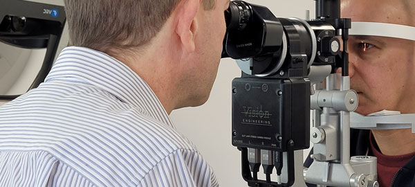
A full HD stereo camera within the optical system of a slit lamp enables stereoscopic viewing through the DRV along with the ability for 3D video/image capture. Captured imagery can be played back in stereo on the DRV or in mono on a regular monitor.
Benefits of the DRV digital stereoscopic slit-lamp imaging system include:
- Ability for an observer to view the examiner’s view in real time in 3D to provide an immersive teaching/training experience
- Parity of the optical image with the digital representation
- Ability to capture images/video in 3D which are more representative of a live examination compared to conventional 2D slit-lamp imaging systems
- Common file saving format allowing for images to be uploaded to image analysis software from other providers which allow for real time analysis/labelling of video/images e.g. to measure lesions or grade redness
- Ability to connect the system to the internet for remote supervision – a remote observer can view the live video feed from the system through a DRV and watch in 3D
- Ergonomic
Slit Lamp Biomicroscopy
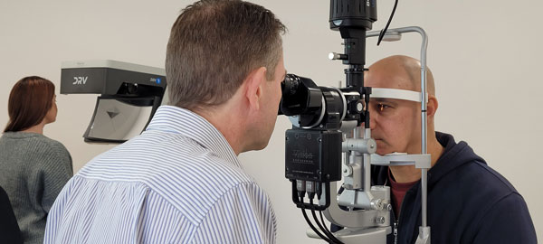
Slit-Lamp biomicroscopy is a difficult skill to learn and to teach. In clinical practice, the presence of a slit-lamp camera system is not common – usual practice may be where the examiner tilts their head to the side and asks the observer/trainee to have a peek through the eye-pieces by which time the patient has moved and the observer/trainee struggles to see what they were intended to.
Slit-lamp cameras have been shown to accelerate the learning curve for developing skills in performing slit-lamp examination, but it is often difficult to capture suitable training material images due to issues with exposure and frame rate.
Where cameras are used, the image captured is not normally 3D so the observer lacks a stereoscopic view as would be seen through the eyepieces.
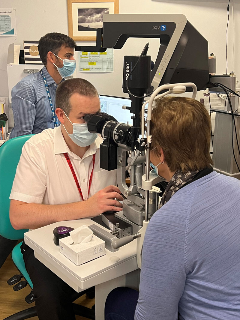
Dan Lindfield, Consultant Ophthalmologist at Royal Surrey County Hospital has been assessing the effectiveness of Vision Engineering’s DRV stereo display as a co-observational and training tool.
It is integrated with my Haag Streit slit lamp using a stereo camera (also manufactured by Vision Engineering) which allows me to use the eye pieces as usual whilst simultaneously presenting a live high definition digital stereo image on the DRV.
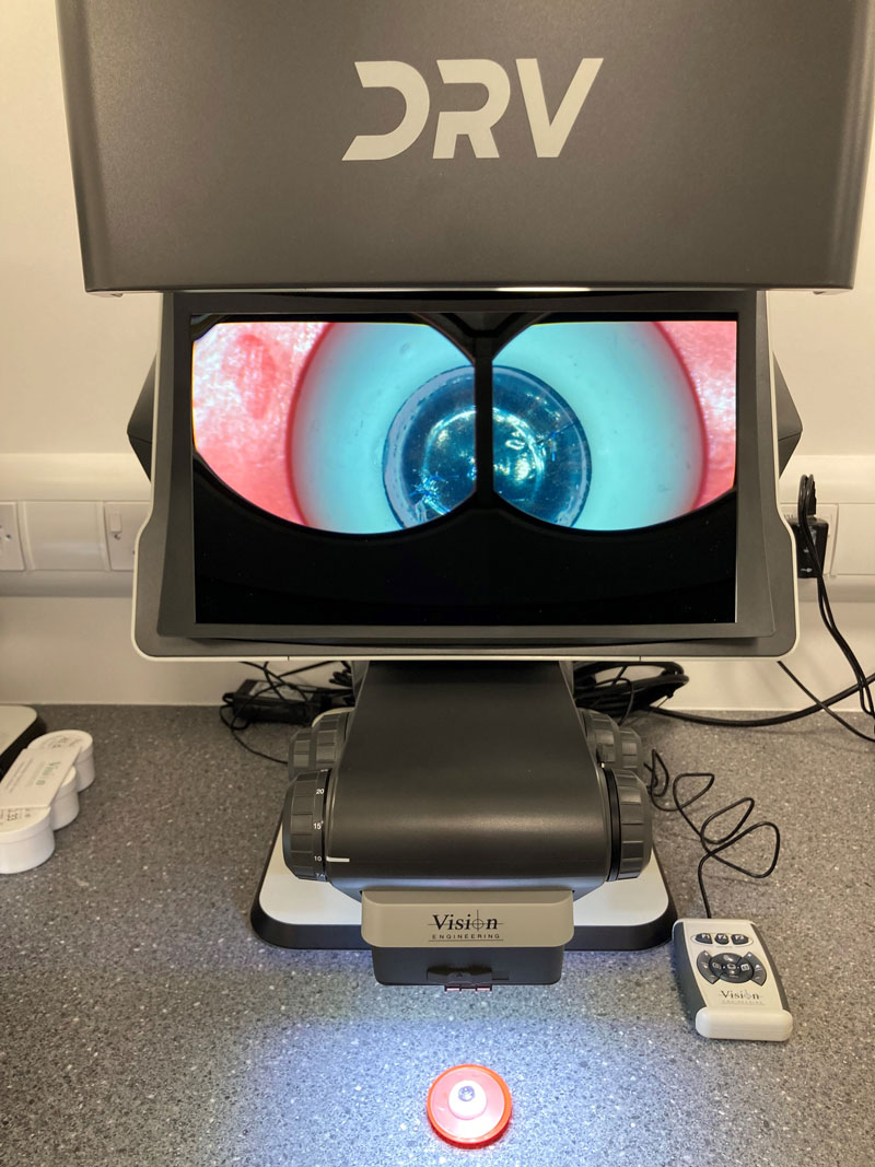
The quality of the DRV stereo image is superb. The high resolution emulates the slit lamp image well and concurrent real-time viewing of clinical findings in true 3D for both trainee and observer is unique. What’s more the observer does not require 3D glasses to view, the DRV image presents a “floating” 3D image in front of the system.
I have found the system very useful in illustrating common and complex pathology to Optometrists, nurses and doctors in training. I foresee the device has great potential for telemedicine / remote decision making in many areas of medicine, not just Ophthalmology. For teaching it allows either real time simultaneous viewing or the ability to record 3D still images and video for later playback and educational review.
Slit Lamp Teaching
In a time where the upskilling of allied health professionals is so necessary to solve a workforce crisis, the use of a 3D slit-lamp imaging system poses a potential solution to provide a high fidelity stereoscopic teaching system to train slit-lamp clinical skills.
The 3D camera system within the slit-lamp can output a stereoscopic video in real time to a DRV. This facilitates high fidelity training opportunities and accelerates the learning curve for clinical skills for junior doctors and allied health professionals.
For optimal stereo imagery with an immersive training experience, the trainee will use the DRV which can output in stereo to multiple DRVs or in mono to a display/projector for presentation to an audience.
Either 2D or 3D videos can be captured and stored for training purposes, specifically useful in the training of identifying unusual and/or rare conditions.
Surgical Simulation Training
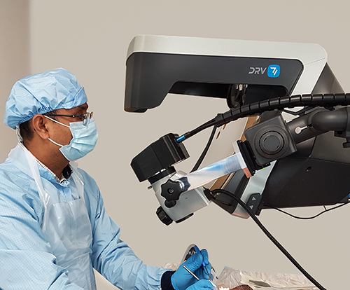
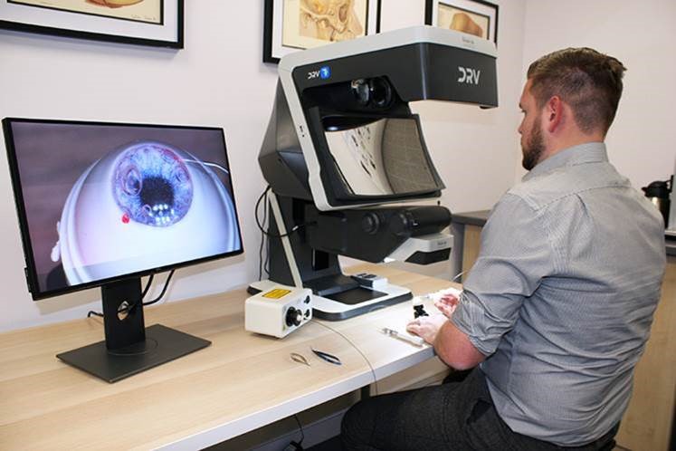
The DRV-Z1 combines a digital stereo display with a microscope which brings a number of benefits over conventional table top microscopes currently in use for surgical simulation training. The ergonomic design of the microscope system improves operator comfort meaning that the user can practice for longer periods whilst remaining comfortable.
Sub-specialty procedures for glaucoma, retina, cataract and corneal surgery can be rehearsed and refined to ensure competence and confidence.
A significant benefit of the DRV-Z1 is that it facilitates high quality video recording. Captured video recordings are of the same view as what was seen by the operator, which differs from conventional table top microscopes where the recorded video often suffers from low refresh rate and disparity of the field of view, focus and colour rendition, when compared to the view that is seen by the operator.
This key feature facilitates high quality supervision which may be done live and in 3D.
Minimally Invasive Glaucoma Surgery (MIGS) Training
The design of the zoom microscope module allows adjustment of the viewing angle which, combined with an engineered holder to mount the model eye, simulates the position and angle of a patient’s eye for trainees to practice their surgical skills, for example trabeculotomy and canaloplasty.
The viewing angle can be adjusted to suit the individual’s and their trainer’s preference.
The ergonomic design of the DRV improves operator comfort allowing the user to practice for longer whilst remaining comfortable.
Surgical: Operating Room Co-observation
A Vision Engineering stereo camera positioned on the microscope in place of the teaching head eyepieces, enables a co-observer to view the surgical procedure on the DRV in stereo without being in close proximity to the patient and surgeon and so allows both primary user and co-observer to view a procedure unobstructed.
Additionally, still shots and video can be saved on a PC for later playback and educational review in stereo.
By connecting DRVs over a network, real-time imagery can be streamed to remote DRVs for sharing with international colleagues.

Surgical Skills Training
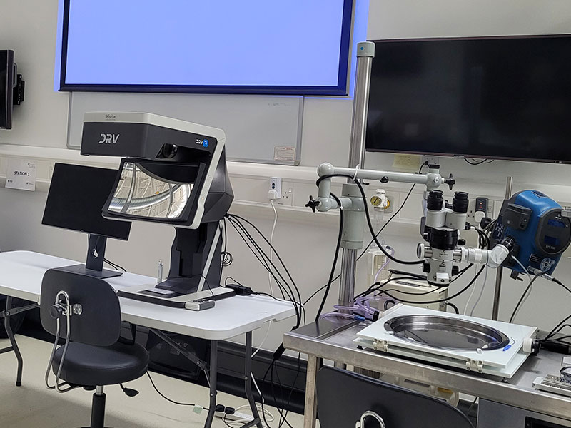
A stereo camera positioned between the microscope optics and eyepieces allows simultaneous viewing of the optical stereo image in the microscope and the digital stereo image in the DRV.
Traditionally, a trainer would supervise the trainee’s performance by viewing a monoscopic image via an uncomfortable teaching tube or by watching a digital image on a mono screen. Not only does this mean the absence of stereo depth within the viewed image, but important information that may only be visible in one channel may be missed if the mono image is displaying the other channel.
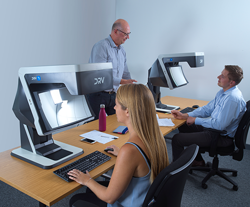
The DRV transforms the teaching capability in clinical skills labs as the trainer can supervise a trainee’s work comfortably in stereo with the benefit of depth perception.
By swapping seats, a trainer can equally demonstrate techniques to the trainee. Multiple DRVs can be connected in series allowing multiple trainees to watch in stereo.
Surgical Teaching
Captured stereo videos can be stored for students and trainees to review on remote DRVs without needing to wait for patients or taking up space in the OR. Pre-recorder stereo videos on specific surgical procedures can be recalled instantly allowing students to complete their studies according to schedule.
Specific sections of videos or still shots can be exported to documents for the preparation of digital and e-learning materials.
Future Capabilities
In the future, it will be possible to use the TriTeQ3 technology for ergonomic stereoscopic heads-up viewing and a remote telemedicine display for consultation and remote diagnosis.
HEADS-UP SURGERY
- Comfortable heads-up surgery
- No need for additional eyewear to view the stereo image
- Can be used whilst wearing PPE
- Stereo cameras allowing :
- Telemedicine
- Remote consultation (diagnostic hubs)
- Digital enhancement
- Capture stereo stills or video
- Retain eyepieces if required
- Teaching (with remote displays)
- Ergonomic
TELEMEDICINE
DRVs can be connected side by side via twin HDMI cables, or streamed across continents over networks, for real-time collaborative analysis of data or remote consultation and Hub & Spoke teaching all in 3D stereo
