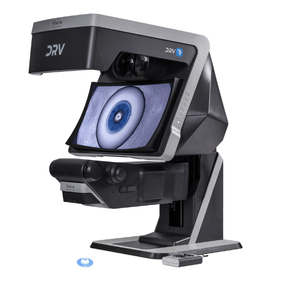
Deep Reality Viewer (DRV) is a unique, glasses-free stereo display designed to provide fully interactive, real time, natural 3D visualisation with outstanding depth perception.
Deep Reality Viewer lets you view subjects in stereoscopic 3D without special glasses or a headset. Our TriTeQ³ technology combines optical and digital design to send two independent optical paths to your eyes. Each eye sees the subject from a slightly different angle, just as in natural stereo vision.

See true 3D depth without glasses
Experience immersive, glasses-free digital stereo 3D with natural depth perception, exceptional image brightness and zero cross-talk, ghosting or flicker. Height-adjustable and precision manufactured in the UK, it enables real-time collaboration and communication for an effortless 3D viewing experience.
Real time collaboration on a global scale
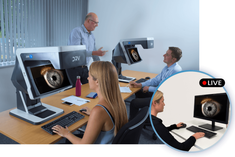
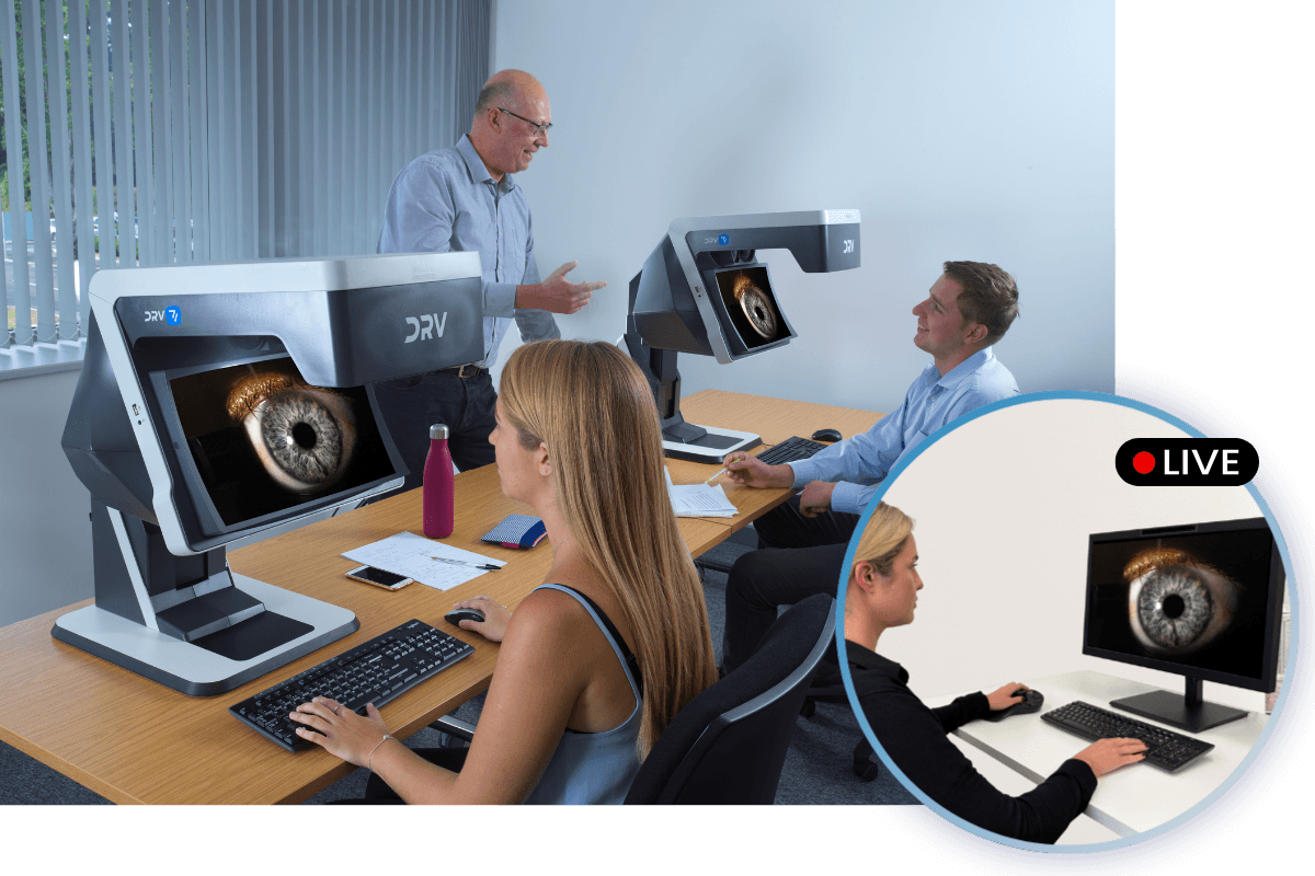
Share 3D full HD images across networks to other DRV systems in real time. Colleagues view live footage remotely, from the operating theatre to a second opinion thousands of miles away, without leaving the room. Record and recall archived 3D images and video easily.

Our design philosophy centres on user ergonomics
DRV’s ergonomic design encourages good body posture with reduced eye fatigue which maximises productivity and quality over long periods of use.
Built for your workflow
DRV Z-1
The DRV Z1 combines the display with a built-in zoom microscope. It works as a complete stand-alone system that’s ready to use out of the box, and it’s designed for simulated surgical training.
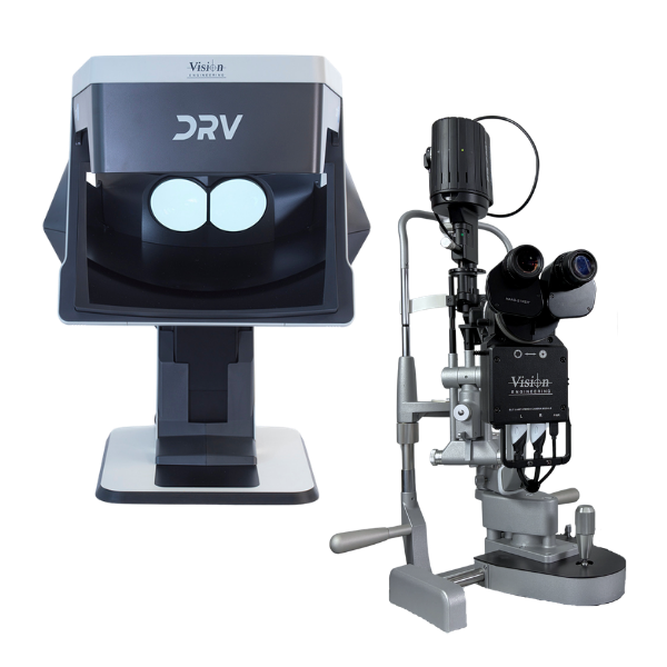
DRV D-1 and Stereo Camera Module
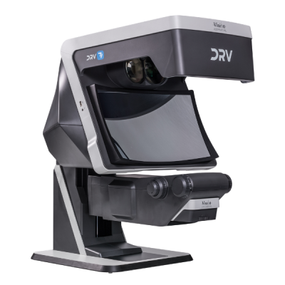
Add our Stereo Camera Module to a compatible slit lamp, stereo microscope or operating microscope, and the DRV D1 shows its image in stereo. You keep the equipment you already have, and you can view, record, play back and share images in 3D.
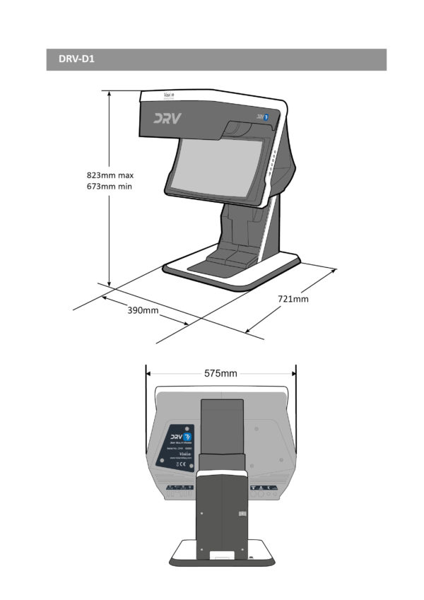
DRV D-1
The DRV D1 also works as a display on its own. You can connect it to a PC, a stereo camera or a stereo endoscope, and play back recorded stereo video. It also supports heads-up surgery with operating microscopes. You sit upright instead of bending over an eyepiece, and you don’t need polarised glasses over your prescription eyewear. You can also link several DRVs so everyone sees the same image in 3D.
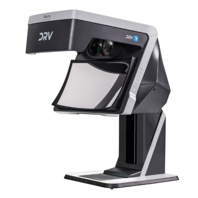
DRV requires minimal setup as the stereo generating optics of DRV are precisely factory aligned which ensures optimal stereo fusion from the outset. No in-use maintenance or adjustment of image alignment is necessary.
DRV for Ophthalmology and ENT
Slit Lamp Imaging System
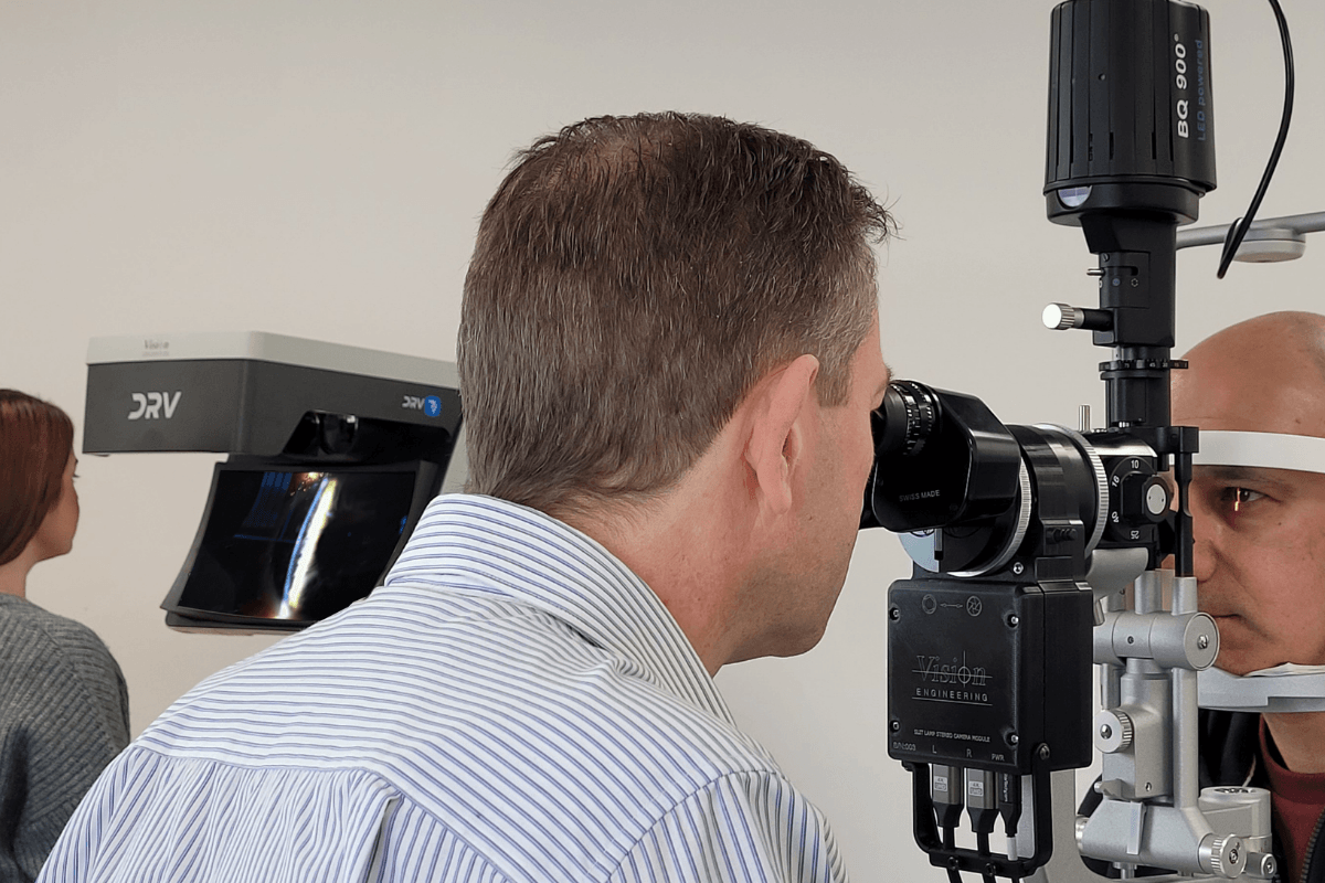
A Full HD stereo camera sits within the slit lamp’s optical system. You view the image through DRV in stereo, glasses-free, and capture 3D video or stills. Play captured imagery back in stereo on DRV, or in mono on a regular monitor.
DRV delivers a widescreen digital stereo 3D image. Connected to the slit lamp, it gives real-time stereoscopic co-observation of the patient examination. Depth and magnification match the optical image.
For training, DRV outputs in stereo to multiple DRVs, or to 2D or 3D monitors for presentations. Observe procedures in real time, or record them in 3D for later review.
Slit Lamp Biomicroscopy
Slit lamp biomicroscopy is a difficult skill to learn and to teach. Slit lamp cameras shorten the learning curve for examination skills. Exposure issues and the lack of a stereoscopic image make useful training material hard to capture. Connect the slit lamp to DRV and observers view or capture the real-time video feed in 3D.
Slit Lamp Teaching
The 3D camera system within the slit lamp outputs stereoscopic video to DRV in real time. Trainees get high-fidelity training and a faster learning curve for clinical skills.
Surgical Training
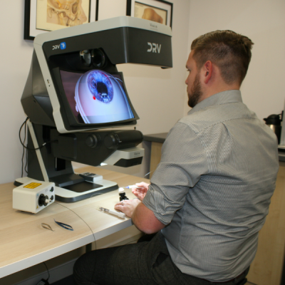
DRV-Z1 combines a digital stereo display with a microscope. Its ergonomic design improves operator comfort, so you practise for longer in the dry lab or wet lab. Rehearse and refine sub-specialty procedures for glaucoma, retina, cataract and corneal surgery.
DRV-Z1 records stereo video of the same view the operator sees. Conventional table-top microscopes often record at a low refresh rate. Their field of view, focus and colour can differ from the operator’s view.
Surgical Skills Training
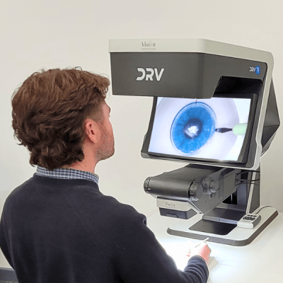
Traditionally, a trainer supervises through a mono teaching tube or on a mono screen. Viewing one channel removes stereo depth and raises the risk of missing information.
With DRV, the trainer supervises in stereo. Swap seats and the trainer demonstrates a technique. Connect multiple DRVs in series for larger training sessions, all with stereo viewing.
Minimally Invasive Glaucoma Surgery Training
The zoom microscope module’s viewing angle adjusts. An engineered holder mounts the model eye. Together they simulate the position and angle of a patient’s eye. Trainees practise surgical skills such as trabeculotomy and canaloplasty.
Tele-Ophthalmology
Connect DRVs side by side with twin HDMI cables, or stream across continents over networks. Every site views in 3D stereo.
Colleagues analyse data together in real time. You can also run remote consultations and hub-and-spoke teaching.
Testimonials
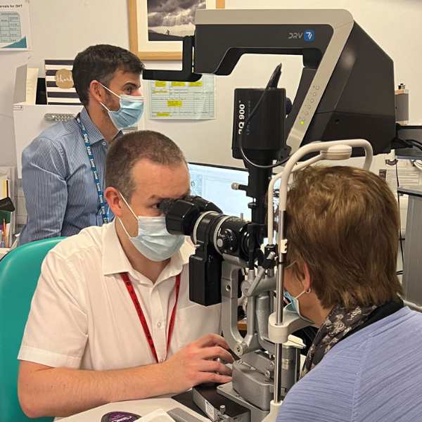
The quality of the DRV stereo image is superb. The high resolution emulates the slit lamp image well and concurrent real-time viewing of clinical findings in true 3D for both trainee and observer is unique. What’s more the observer does not require 3D glasses to view, the DRV image presents a “floating” 3D image in front of the system.
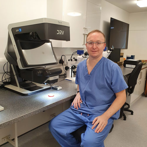
Through the use of the DRV-MZ1 trainees in the Glasgow based surgical simulation suite are reaping the dual benefits of optical stereo microscopy and advanced digital technology in a single system. The DRV-MZ1 provides impressive levels of simulation by delivering fully immersive 3D visualisations with outstanding perception of depth.
Technical information
| Category | Specification | Detail |
| Display Head | Resolution | 1920 x 1080 per channel |
| Image size on concave mirror | 400 x 225 mm in 16:9 aspect ratio | |
| Digital zoom | 2x | |
| Working distance (maximum) | 182 mm | |
| Inputs | Power supply | 100–240 VAC 50/60 Hz |
| Outputs | Image capture | USB 2.0 |
| Video capture | HDMI cable to an external video capture card | |
| Connection to external 2D/3D monitor | HDMI | |
| Connection to second or multiple DRVs | HDMI daisy chain / Wi-Fi connection | |
| Stand | Counterbalanced stand | 150 mm vertical travel |
| Fully adjustable sub-stage illumination | Optional | |
| Zoom Module | Zoom module | 10:1 optical zoom with fully adjustable surface illumination |
| Weight | Maximum system weight | 45 kg |
| Objective Lens | Zoom Range | Working Distance ‘A’ | Field of View at MAX zoom | Field of View at MIN. zoom |
| 0.33x | 6.1x – 61x | 182 mm | 6.5 mm / 3.7 mm | 65 mm / 37 mm |
| 0.4x | 7.4x – 74x | 138 mm | 5.4 mm / 3.0 mm | 54 mm / 30 mm |
| 0.5x | 9.3x – 93x | 93 mm | 4.3 mm / 2.4 mm | 43 mm / 24 mm |
| Stereo Camera Module | |
| Image Sensors | 1 / 2.8” |
| Channel Resolution | 1920 x 1080 (per channel) |
| Pixel Size | 2.9 µm |
| Colour Depth | 8 bit |
| Power Source | 5 V via USB |
| Power Consumption | 4.9 W |
| Video Transmission Method | 2 x HDMI |
| Frame Rate | 60 fps |
| Aperture Control | Yes |
| Beam Splitter | Fixed and selectable options |
| Power and Video Ports | Located on bottom of Camera 234 |
| Dimensions (L x W x D) | 234 x 120 x 80 mm |
Get the full DRV brochure. Fill in your details below.
Frequently asked questions
Below you can find some of the most common questions.
Yes. DRV can be interfaced with external digital stereo cameras, digital stereo endoscopes, digital stereo microscopes, etc via two video outputs, one for the left eye and one for the right eye. DRV also supports HDMI stereo frame packing method over a single HDMI cable. Other stereo video formats can be also supported using an auxiliary PC between the capturing device and the DRV.
Yes. Many popular Common Main Objective (CMO) stereo microscopes can be interfaced with the DRV, using a DRV stereo camera module available separately.
Yes, using two HDMI cables (one for the left channel and one for the right channel) and an adequate graphics card, DRV can be connected to the PC and can be used as a stereo display for viewing in true stereo computer generated 3D models or stereo videos/photos.
DRV doesn’t impose additional requirements beside those required by the stereo software package in use. For software packages which require OpenGL quad buffered stereo support, any quad buffered stereo capable graphics card (NVIDIA Quadro or AMD RadeonPro/FirePro) can be used with DRV. For software packages that operate in dual screen mode, any graphics card capable to support two video outputs can be used with DRV.
Active shutter glasses technology, suffers from flickering, cross-talk and ghosting, which provokes eye strain. DRV delivers a brighter image with no flickering or ghosting. NVIDIA “3D Vision” active shutter glasses and emitter are no longer supported by the manufacturer.
For applications where stereo 3D viewing is required (without head tracking), DRV delivers a bright, high resolution stereo image at a comfortable viewing distance. DRV doesn’t suffer from the problems associated with the use of VR goggles, such as, screen door effect, VR motion sickness, isolation, eye-strain, “welders neck”, hygiene. The advantage of not having to wear VR goggles also means the user can comfortably wear their prescription glasses, if needed, and retain their peripheral vision for easy reference to documents, instruments and colleagues.
Yes. DRV allows you to use your prescription glasses and eliminates the need to wear additional polarisation glasses.
No. DRV is a stereo display which works out of the box. We also supply easy to follow documentation on NVIDIA and AMD graphics card configurations, as well as guidance on interfacing DRV with external image capturing devices.
No. It takes approximately 15 minutes to unpack and assemble.
DRV requires minimal setup as the stereo generating optics of DRV are precisely factory aligned which ensures optimal stereo fusion from the outset. No in-use maintenance or adjustment of image alignment is necessary.
DRV is supplied with a comprehensive user guide. Unpacking, assembly and handling instruction videos are also available.