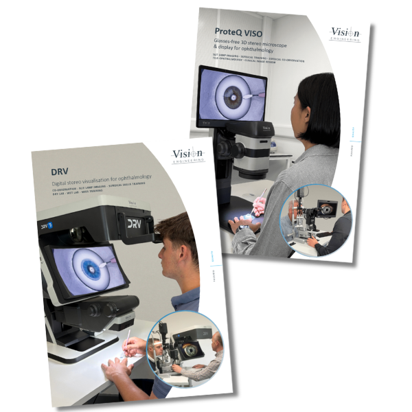
In surgical and ophthalmic disciplines, co-observation, teaching and training are typically performed using optical stereo microscopes with mono teaching tubes, or digital cameras with mono screens. Both strip out the depth that makes clinical detail clear.
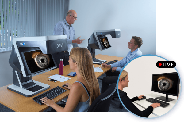
Vision Engineering’s glasses-free 3D heads-up display technologies create new capabilities. You view stereo images from an integrated microscope, or interface with existing optical microscopes and slit lamps, with no glasses or headset. Digital stereo images can be viewed locally or remotely, in real time or played back from a recording.
Our glasses-free 3D displays
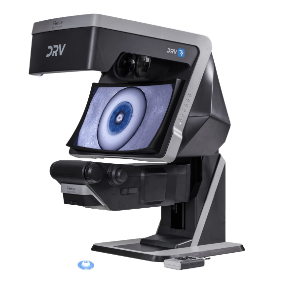
DRV
The highest depth perception, for advanced surgical and diagnostic work.
The patented “TriTeQ³” technology behind DRV’s stereo image presentation overcomes the need for polarised glasses by projecting independent optical channels to each eye, which replicates our natural stereo vision and perception of depth.
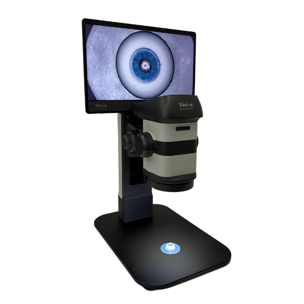
ProteQ VISO
A compact flat panel designed to integrate seamlessly into modern healthcare workspaces
Using advanced lenticular display technology with integrated eye tracking, the ProteQ VISO dynamically adjusts the viewing zones to maintain optimal stereo perception. The output is stable, flicker-free, stereoscopic depth without the need for polarised glasses.
Depth perception without the eyepieces
A conventional operating microscope fixes your head to the eyepieces for hours. Teaching tubes show mono. 2D monitors flatten the anatomy. Polarised 3D glasses add extra eyewear and can be awkward if you already wear prescription glasses.
Vision Engineering’s heads-up technology frees you from the eyepieces. With your posture upright, you move your head freely and maintain natural hand-eye coordination. Most surgeons using a heads-up display report better posture and less neck strain than conventional microscopy.
A glasses-free 3D heads-up display goes one step further. Two full HD channels combine to put a high resolution stereo image of the surgical field on screen, with nothing on your face. Image quality and depth stay intact. If you already wear prescription or safety glasses, the display still works for you. Training takes very little time, because you view the anatomy in stereo, the way you always have.
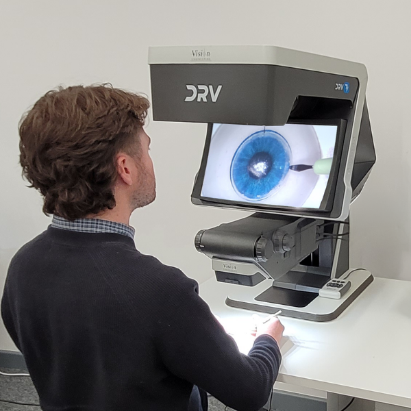
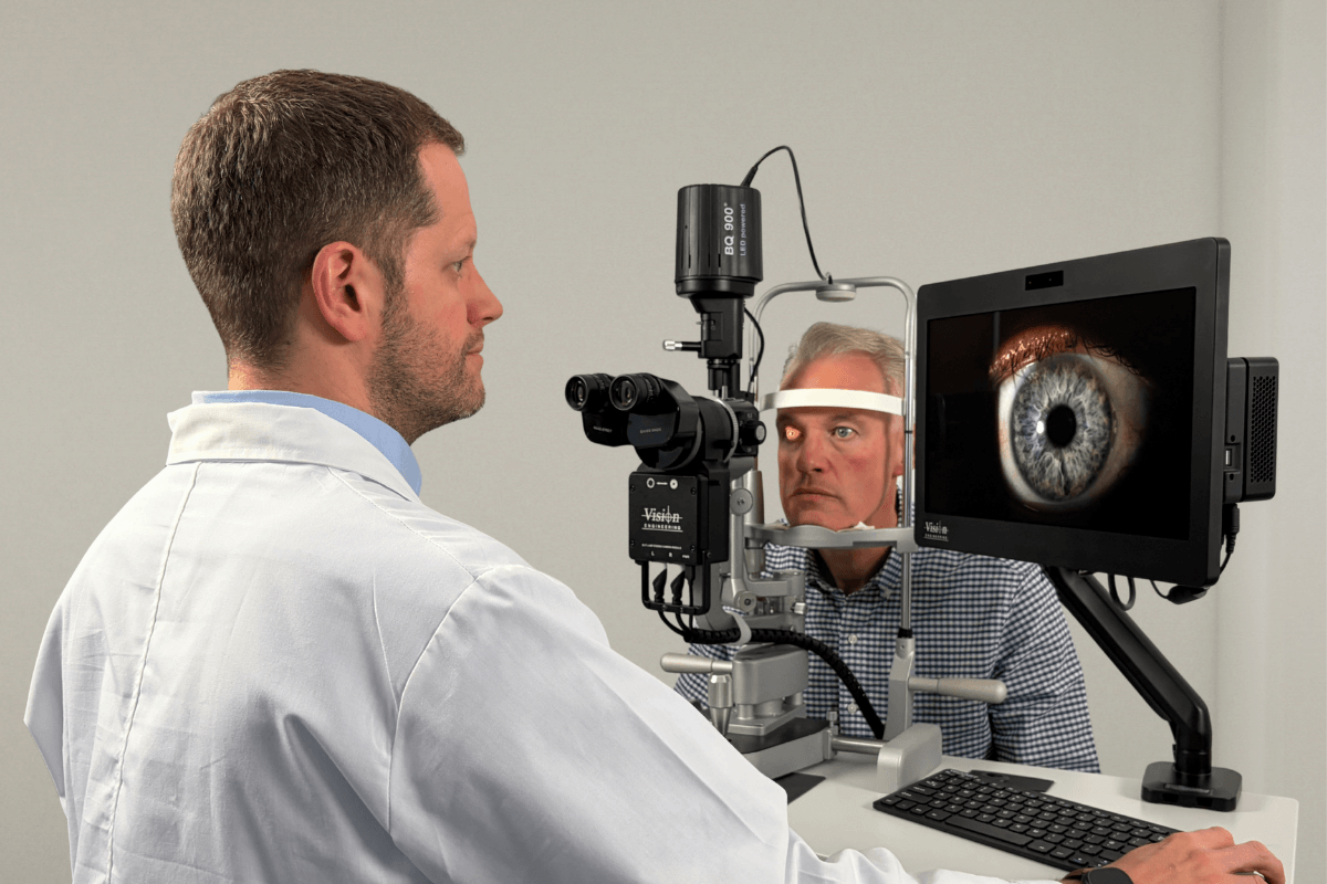
3D Slit Lamp imaging system
A full HD stereo camera fits within the optical system of a slit lamp, so you view a live stereoscopic image on the display and capture 3D video and stills. Captured imagery plays back in stereo on the display, or in mono on a regular monitor.
Benefits of the glasses-free slit lamp imaging system:
- An observer views the examiner’s view in real time in 3D, for an immersive teaching experience
- Parity of the optical image with the digital representation
- 3D image and video capture more representative of a live examination than conventional 2D slit lamp imaging
- A common file format, so images upload to third-party analysis software for real-time labelling, lesion measurement or redness grading
- Internet connection for remote supervision, so a remote observer watches the live feed in 3D
Slit Lamp biomicroscopy
Slit lamp biomicroscopy is difficult to learn and to teach. In clinical practice, a slit lamp camera is not common. Usual practice is the examiner tilting their head aside and asking the trainee to take a peek through the eyepieces, by which time the patient has moved and the trainee struggles to see what was intended.
Slit lamp cameras have been shown to accelerate the learning curve for examination skills, but suitable training images are often hard to capture due to exposure and frame rate. Where cameras are used, the image is not normally 3D, so the observer lacks the stereoscopic view seen through the eyepieces.
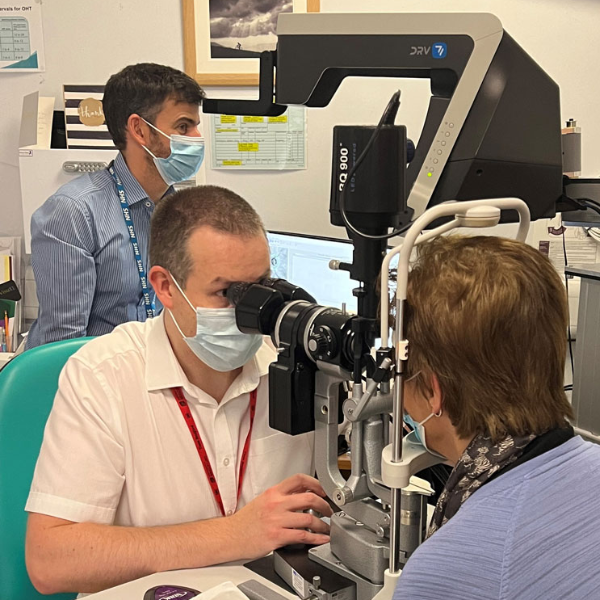
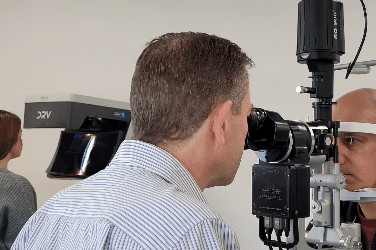
Slit Lamp teaching
Upskilling allied health professionals matters for the workforce. A 3D slit lamp imaging system offers a high-fidelity stereoscopic way to train slit lamp clinical skills.
The 3D camera in the slit lamp outputs stereoscopic video in real time to the display. This accelerates the learning curve for junior doctors and allied health professionals. For presentation, the system outputs in stereo to multiple displays, or in mono to a screen or projector for an audience. 2D or 3D video can be captured and stored, useful for training on unusual or rare conditions.
See Slit Lamp imaging using the DRV → or with the ProteQ VISO →
Surgical simulation training
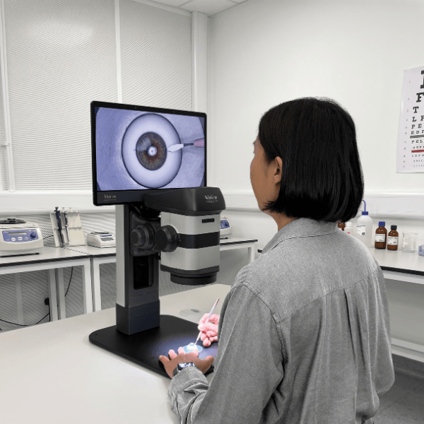
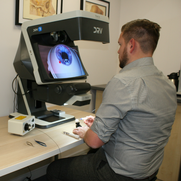
A digital stereo display combined with a microscope brings benefits over conventional table-top microscopes used for surgical simulation. The ergonomic design improves operator comfort, so users practice for longer.
Sub-specialty procedures for glaucoma, retina, cataract and corneal surgery can be rehearsed and refined.
Video recording captures the same view the operator sees. Conventional table-top microscopes often record video that suffers from low refresh rate and disparity of field of view, focus and colour against the operator’s view. Matched-view recording supports high-quality supervision, live and in 3D.
Minimally invasive glaucoma surgery (MIGS) training
The zoom microscope module adjusts its viewing angle. Combined with an engineered holder to mount the model eye, it simulates the position and angle of a patient’s eye, so trainees practice trabeculotomy and canaloplasty. The angle adjusts to suit the trainee and trainer. The ergonomic design lets users practice for longer in comfort.
See Surgical simulation training using the DRV →
or with the ProteQ VISO →
Surgical: operating room co-observation
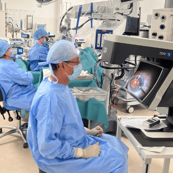
A stereo camera positioned on the surgical microscope, in place of the teaching head eyepieces, lets a co-observer view the procedure on the display in stereo without being close to the patient and surgeon. Both primary user and co-observer view the procedure unobstructed.
Stills and video save to a PC for later playback and educational review in stereo. Connected over a network, real-time imagery streams to remote displays for sharing with international colleagues.
Surgical skills training
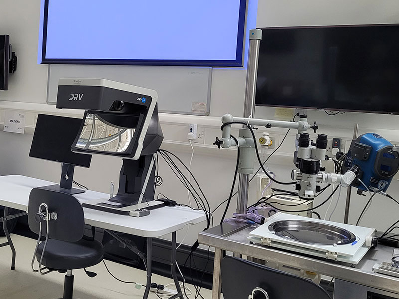
A stereo camera between the microscope optics and eyepieces allows simultaneous viewing of the optical stereo image in the microscope and the digital stereo image on the display.
Traditionally a trainer supervises via an uncomfortable mono teaching tube or a mono screen. Both removes stereo depth, and detail visible in only one channel can be missed if the mono image shows the other channel. Stereo supervision keeps the depth, so the trainer sees a trainee’s work in full.
By swapping seats, a trainer demonstrates techniques. 2D or 3D displays can be connected for audience reviewing.
Surgical teaching
Captured stereo videos stored for students and trainees to review on remote displays, without waiting for patients or taking up space in the OR. Pre-recorded stereo videos on specific procedures recall instantly, so students study to schedule. Specific sections or stills export to documents for digital and e-learning materials.
See Surgical training using the DRV → or with the ProteQ VISO →
Future Capabilities
In the future, it will be possible to use the TriTeQ3 technology for ergonomic stereoscopic heads-up viewing and a remote telemedicine displays for consultation and remote diagnosis.
Heads-up surgery
The glasses-free display supports comfortable heads-up surgery, with no additional eyewear to view the stereo image, usable while wearing PPE. Stereo cameras allow digital enhancement and the capture of stereo stills or video. Eyepieces are retained if required.
Telemedicine
Displays connect side by side via twin HDMI cables, or stream across continents over networks, for real-time collaborative analysis, remote consultation and hub-and-spoke teaching. All in 3D stereo. This supports remote consultation through diagnostic hubs and teaching with remote displays.
What consultant ophthalmologists say
“Through the use of the DRV-MZ1 trainees in the Glasgow based surgical simulation suite are reaping the dual benefits of optical stereo microscopy and advanced digital technology in a single system. The DRV-MZ1 provides impressive levels of simulation by delivering fully immersive 3D visualisations with outstanding perception of depth.”
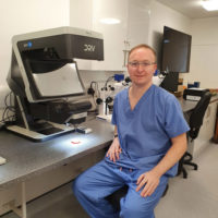 David Lockington, Consultant Ophthalmologist, Glasgow
“It is integrated with my Haag Streit slit lamp using a stereo camera (also manufactured by Vision Engineering) which allows me to use the eye pieces as usual whilst simultaneously presenting a live high definition digital stereo image on the DRV.
The quality of the DRV stereo image is superb. The high resolution emulates the slit lamp image well and concurrent real-time viewing of clinical findings in true 3D for both trainee and observer is unique. What’s more the observer does not require 3D glasses to view, the DRV image presents a “floating” 3D image in front of the system.
I have found the system very useful in illustrating common and complex pathology to Optometrists, nurses and doctors in training. I foresee the device has great potential for telemedicine / remote decision making in many areas of medicine, not just Ophthalmology. For teaching it allows either real time simultaneous viewing or the ability to record 3D still images and video for later playback and educational review.”
Dan Lindfield, Consultant Ophthalmologist, Royal SurreyBUILT ON 65+ YEARS OF MICROSCOPY AND 3D VISUALISATION
1958
Vision Engineering founded
2020
Queen’s Award for Enterprise
ISO 9001 & ISO 13485
certified
ISO 17025
UKAS accredited
See it in your clinic
Request a demonstration and download the ophthalmology brochure below. We’ll set the system up with your slit lamp or microscope so you can judge the stereo image yourself.