Ophthalmology and ENT
In surgical and ophthalmic disciplines, co-observation, teaching and training are typically performed using optical stereo microscopes with mono teaching tubes or digital cameras with mono screens.
Vision Engineering’s unique glasses-free TriTeQ3 technology creates new capabilities allowing users to comfortably view stereo images from the integrated microscope or interfacing with existing optical microscopes or slit lamps. Digital stereo images can be viewed locally or remotely in realtime or aftertime by watching recorded content. FIND OUT MORE.

MRI / CT Scan Imaging
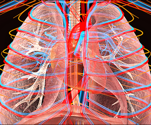
Medical professionals need to look at ever more complex and increasing volumes of imaging data to make diagnoses and plan surgical treatments.
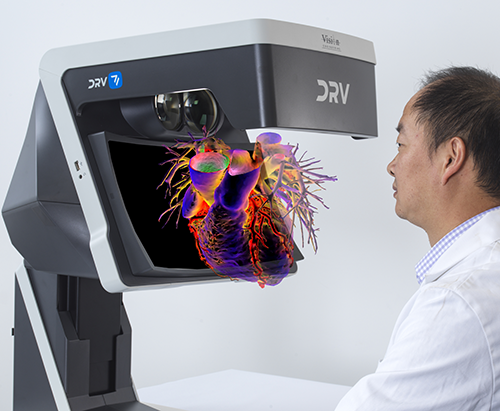
DICOM data sets generated from MRI/CT equipment have traditionally been viewed as slices on mono screens using a technique known as multi-planar reconstruction. However, advances in volume rendering software has created the ability to view the DICOM data set in 3D. DRV takes visualisation to the next level by enabling the specialist to view the data in stereo 3D which adds the benefit of natural depth perception.
Education, Training and Presentation
DRV can be used as a teaching tool in medical environments and universities. The ability to view educational material in 3 dimensions that make use of the stereo cues and understanding of geometry offers a unique benefit to today’s students.
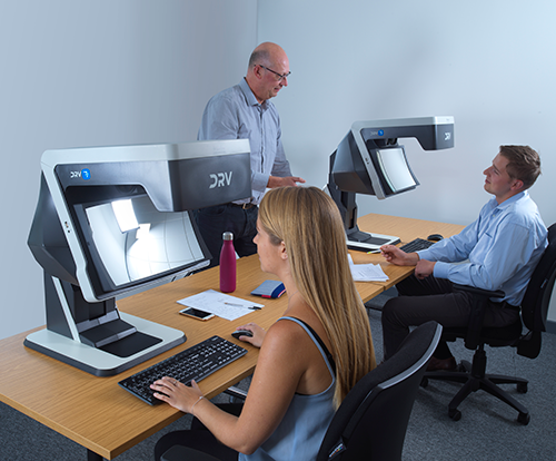
Moreover, the ability to connect multiple DRVs can be explored in a teaching environment, whether it be in person or remote. Students can follow precisely what the teacher is presenting in stereo 3D and, equally, the teacher can accurately assess a student’s work.
Stereo Microscope Integration
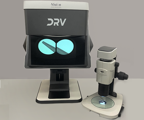
DRV is a breakthrough in the field of digital stereo microscopy. Optical stereo microscopes can be enhanced by the integration of a digital stereo camera module and the DRV display.
This allows high quality images from the host microscope to be viewed, captured and shared in stereo, enabling improved collaboration locally and internationally.
Health issues associated with binocular eye-piece microscopes are already well understood. DRV’s viewing ergonomics eliminate these issues, and allow the user to maintain comfortable posture and wear their prescriptive eyewear if required. This improves productivity and accuracy over long working periods.
Uniquely, the ability to separate the DRV stereo display from the microscope and view remotely, keeps the user protected and safe whilst the microscope may be placed in hazardous areas.
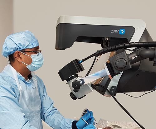
Operating Microscope Integration
Over recent years, attempts have been made to achieve a heads-up imaging solution in surgical applications to help surgeons overcome long-term postural issues and eye strain. However, current solutions invariably rely on the use of active or passive polarisation glasses which, besides being awkward to wear in a surgical environment (especially if prescriptive eyewear is also required), are well known for deterioration of the image quality.
Using a stereo camera interface, the DRV can be integrated with medical microscopes to provide glasses-free, heads-up stereo viewing without compromising the image quality. The improved ergonomics, avoidance of additional eyewear and the ability to view, capture and share live images across networks all helps to improve workflows, efficiencies and patient outcomes.
Geospatial
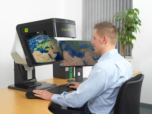
GIS professionals work intensively and for long periods which can be stressful on the body and the eyes. At the same time, GIS professionals must be able to interpret images from many sources of varying quality. Using new, patented digital technology, CONTOUR optimizes image quality, depth and ergonomics.
CONTOUR next generation display delivers a unique high quality combination of image clarity, brightness, consistency, ease of use, and operator comfort. CONTOUR’S 3D full high definition (3D FHD) image provides crisp stereo imagery in a unique, glasses-free, system that ensures the image is delivered precisely, consistently, and accurately, with no flickering, ghosting, or cross-talk.
Mechanical Engineering
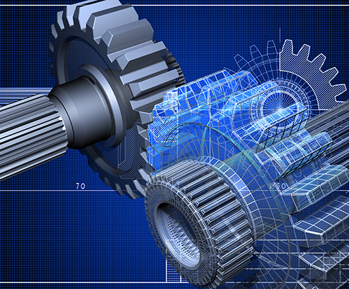
Engineers traditionally view their 3D rendered designs on 2D monitors which creates a need to predict the missing depth and volumetric information.
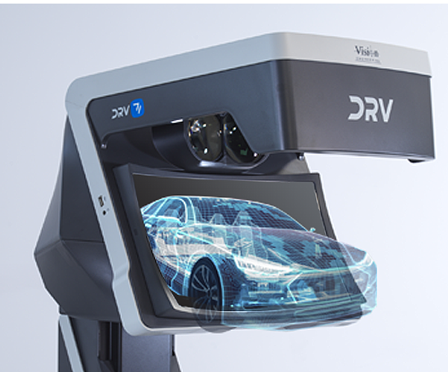
When used with 3D CAD design software, the DRV allows engineers to view complex projects with certainty by taking advantage of the DRV’s unique 3D image presentation and the resulting stereo cues.
Virtual Product Development (VPD) & Prototyping
DRV display technology enhances the VPD workflow with the ability to view digital representation of the product in comfortable true stereo 3D, without the need for glasses or awkward headsets.
The stereo cues and natural depth perception created by the DRV’s unique stereo image allows designers to preview as close to reality as is possible a product’s final appearance without needing to create functional prototypes.
Telemedicine
Remote diagnoses rely heavily on the ability to view detailed imagery of the patient’s condition. The more detailed the imagery, the more accurate the diagnosis. Typically, saved images are captured and reviewed in 2D format on a computer monitor. Whilst the ability to view 3D images have emerged more recently, it relies on the physician using active or passive glasses which suffer from the well-documented issues of flickering, ghosting and cross-talk which all reduce the image quality.
DRV’s unique stereoscopic image presentation technology overcomes these issues and provides the physician with bright, high-resolution, glasses-free, viewing of the patient’s images with a natural perception of depth.

Remote Viewing
Images acquired from stereo cameras, MRI/CT scans or stereo capable software can not only be viewed in 3D by the local user but, thanks to the DRV’s multi-connectivity capability, images can be shared across the globe so remote colleagues can view precisely the same data, in stereo, in real-time, on their DRV.
The ability to simultaneously share high resolution, stereoscopic images advances the quality of student training, distanced consultation and remote collaboration.
Architecture and Building Information Modelling (BIM)
It is now common practice to wear VR headsets to demonstrate architectural designs and walk-throughs to investors and customers.
Unlike VR headsets which are known to cause issues of isolation, disorientation and motion sickness, DRV provides users a pseudo immersive experience whilst retaining their peripheral vision and sensory stimuli.

VR Game Development
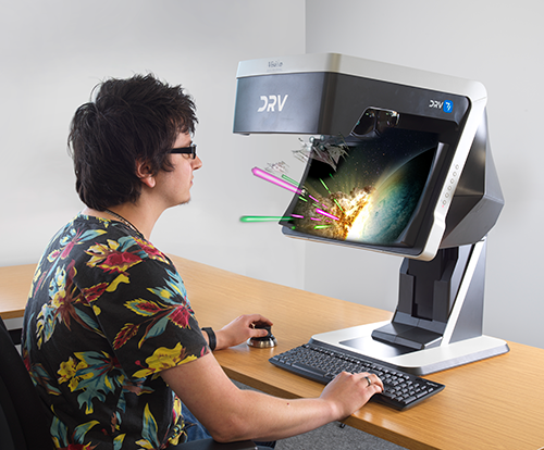
The DRV is the next level in VR game design offering developers the ability to design and collaborate in digital stereo 3D without the need to constantly put on and remove headsets to assess their work,which quickly causes eye fatigue. The immediacy of assessing content, both ergonomically and accurately, adds a faster more comfortable solution for game development.